Breast Cancer HER2 Subtype Identification
Predicting the state of HER2 using Histopathological images
The Customer
The Dow University of Health Sciences (initials: DUHS), is one of the oldest public sector universities in Pakistan. The university comprises of two leading health sciences undergraduate research institutes: Dow International Medical College and Dow Medical College. The University also has a very strong department of Postgraduate studies which monitors various basic medical sciences and clinical sciences programs at DUHS. Established in 1945, it is known for its strong emphasis on economics biomedical, health, and medical research programs. The institution offers undergraduate, post-graduate and doctoral programs in almost all academic disciplines relating to medical sciences.
The Problem And The Solution
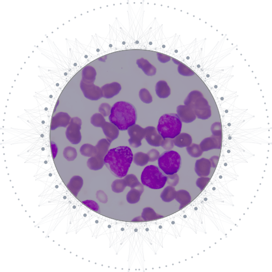
Breast cancer is the leading cause of mortality among women. Every fifth breast cancer patient is diagnosed with HER2 positive breast cancer, which is a very aggressive type of cancer and thus requires timely and accurate diagnosis to limit the chances of mortality. Fluorescence in Situ Hybridization (FISH) test is the most commonly used method for the detection of HER2 Positive breast cancer. However, the process is quite tedious, consumes time and cannot be performed without highly trained practitioners. The solution we developed provided the client a computer aided assistance system that allows practitioners to perform the test more quickly and accurately while enabling them to digitize and store the images for later usage. The FISH test is used as a conventional method in order to manually count the number of HER2 and CEP17 genes in individual cells. To make this process more effective, our system also offered an automated pipeline for cell segmentation and spot counting.
Key Features Of Our Solution
Contrast Enhancement
The contrast enhancement technique plays a vital role in image processing as it brings out the information that exists within the low dynamic ranges of a gray level image. To improve the quality of an image, it enables the performance of operations like contrast enhancement and reduction or removal of noise.

Image Binarization
Binarization is the process of converting a pixel image to a binary image, it is the basis of segmentation. In the old days binarization was important for sending faxes, today it is used for digitizing text or segmentation. The threshold can either be set fixed or adaptive, using a clustering algorithm; this algorithm is called ISO Data Algorithm - it first counts the appearance of each tone in the image and then tries to find a good center.
Cell segmentation
The result of image segmentation is a set of segments that collectively covers the entire image, or a set of contours extracted from the image. Each of the pixels in a region are similar with respect to some characteristic or computed property, such as color, intensity, or texture.
Spot Counting
Each spot represents the 'cytokine signature' of a single cell. Due to diffusion properties, a true spot has a densely colored center which fades toward the edges; the size and/or color intensity of the spots is determined by the amount of cytokine released.
Thanks to Folio3 and our partner Fidel AI’s expert team for developing solution of Breast Cancer HER2 identification. Our client has been able to substantially improve the efficiency and accuracy of their HER2 identification process while enabling the practitioners to digitize and store the images for later usage.
Technologies used: Scikit-image, Matlab+Java for the application.


